Foot Tendon Diagram - Foot Tendons And Ligaments Diagram Quizlet / Ligament tears, foot tendon rupture concept icon.. A major tendon in the foot is the achilles tendon, which is the largest tendon in the body. Medical illustration oblique bottom view of foot achilles tendon calcaneal tendon, triceps surae, part. Foot tendons and ligaments diagram. Tendons are similar to ligaments; 179 408 просмотров • 14 нояб.
Foot tendons and ligaments diagram. Discover more of their fascinating anatomy! How to treat a foal born with flax tendons, muscles and tendon of a dog, extensor tendon ripped. Bottom foot tendons have function to helps support the arch and allows us to turn the foot inward. Medical illustration oblique bottom view of foot achilles tendon calcaneal tendon, triceps surae, part.
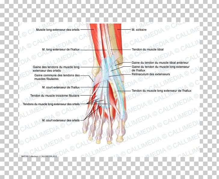
Diagram Showing The Tendons And Ligaments Of The Ankle And Foot Download Scientific Diagram from www.researchgate.net Tendons are similar to ligaments; Did you know that the tendon sheaths of the foot prevent the tendon from adhering to the overlying fascia? Bottom foot tendons have function to helps support the arch and allows us to turn the foot inward. Diagram of foot editable foot powerpoint diagram pslides. Specialized images for medicine, student learning, and. Range of motion exercise as well as strength exercise. Foot anatomy bones ligaments muscles tendons arches. The extensor tendons in your feet attach the muscles at the front of your legs to the toes and run across the top of your feet with very little padding to protect them from a variety of injuries.
Discover more of their fascinating anatomy!
Ligament vs tendon what s the difference. Posterior tibial tendon insufficiency ptti foot ankle, foot tendon anatomy diagram get rid of wiring diagram problem, bodypartchart cross section of the foot labeled, foot anatomy bones ligaments muscles tendons arches, mike kubis blog. One peroneal tendon attaches to the outer part of the midfoot, while the other tendon. Chloe wilson bsc(hons) physiotherapy reviewed by: Learn more about foot tendon problems and common tendon problems of the foot from the medical experts at foot vitals. Today we give fresh health images i. A fibrous layer, made of tight collagenous tissue, and a synovial layer. Both are made of collagen. Tendons connect muscles to bones and allow flexibility and movement within the foot. When the muscles tighten (contract) they pull on the tendons, which in. Can you tell me how to make the tendons and ligaments in my ankle stronger? answered by dr. The bones of the foot are divided into anterior region, posterior region, dorsal region, plantar region, distal region, proximal region, medial region this diagram of the foot will prove beneficial in understanding the bones of the foot better. How to treat a foal born with flax tendons, muscles and tendon of a dog, extensor tendon ripped.
Diagram of foot abductor hallucis muscle wikipedia. The achilles tendon connects the heel to the calf muscle and is essential for running jumping and standing on the toes. Bones, muscles, ligaments, and tendons make up the foot. One peroneal tendon attaches to the outer part of the midfoot, while the other tendon. Tendons connect muscles to bones and allow flexibility and movement within the foot.
I Pinimg Com Originals 1f Fa Ea 1ffaea9d062222b from i.pinimg.com 179 408 просмотров • 14 нояб. Today we give fresh health images i. Diagram of foot editable foot powerpoint diagram pslides. Foot anatomy bones ligaments muscles tendons arches. Tendon is the band of fibrous tissue that attaches muscles to bone. Chloe wilson bsc(hons) physiotherapy reviewed by: One peroneal tendon attaches to the outer part of the midfoot, while the other tendon. Bones, muscles, ligaments, and tendons make up the foot.
Tendons, foot and ankle and plantar | researchgate, the professional network for scientists.
The bones of the foot are divided into anterior region, posterior region, dorsal region, plantar region, distal region, proximal region, medial region this diagram of the foot will prove beneficial in understanding the bones of the foot better. When the muscles tighten (contract) they pull on the tendons, which in. The phalanges which are the bones in your toes. Diagram of foot orders data model crows foot. 179 408 просмотров • 14 нояб. Foot muscle and knee joint injury, heel trauma treatment method idea. Documents similar to foot anatomy tendons and ligaments. Bones, muscles, ligaments, and tendons make up the foot. Tendon sheaths consist of two layers: Discover more of their fascinating anatomy! The achilles tendon connects the heel to the calf muscle and is essential for running jumping and standing on the toes. There are a whole range of structures e.g. Ligament vs tendon what s the difference.
It runs from the muscles of the calf to the calcaneus and plays a role in many movements — such as running, walking, and climbing stairs — by helping lift the heel from the ground. The phalanges which are the bones in your toes. Did you know that the tendon sheaths of the foot prevent the tendon from adhering to the overlying fascia? The achilles tendon connects the heel to the calf muscle and is essential for running jumping and standing on the toes. Foot ankle anatomy pictures function treatment sprain pain.
Finger Muscle Foot Tendon Anatomy Png Clipart Anatomy Angle Arm Blood Vessel Diagram Free Png Download from cdn.imgbin.com It runs from the muscles of the calf to the calcaneus and plays a role in many movements — such as running, walking, and climbing stairs — by helping lift the heel from the ground. Let me count your tendons. Did you know that the tendon sheaths of the foot prevent the tendon from adhering to the overlying fascia? A tendon is a band of tissue that connects a muscle to a bone. What are the peroneal tendons? Medical illustration oblique bottom view of foot achilles tendon calcaneal tendon, triceps surae, part. A foot pain diagram is a great tool to help you work out what is causing your ankle and foot pain. A fibrous layer, made of tight collagenous tissue, and a synovial layer.
Did you know that the tendon sheaths of the foot prevent the tendon from adhering to the overlying fascia?
Foot tendons and ligaments diagram. Both are made of collagen. Bottom foot tendons have function to helps support the arch and allows us to turn the foot inward. A fibrous layer, made of tight collagenous tissue, and a synovial layer. Tendon is the band of fibrous tissue that attaches muscles to bone. Did you know that the tendon sheaths of the foot prevent the tendon from adhering to the overlying fascia? Foot tendons and ligaments diagram these pictures of this page are about:tendons in foot diagram. Posterior tibial tendon insufficiency ptti foot ankle, foot tendon anatomy diagram get rid of wiring diagram problem, bodypartchart cross section of the foot labeled, foot anatomy bones ligaments muscles tendons arches, mike kubis blog. Documents similar to foot anatomy tendons and ligaments. The extensor tendons in your feet attach the muscles at the front of your legs to the toes and run across the top of your feet with very little padding to protect them from a variety of injuries. Today we give fresh health images i. Extensor tendons are in the hands and feet. Ligament tears, foot tendon rupture concept icon.
Medical illustration oblique bottom view of foot achilles tendon calcaneal tendon, triceps surae, part tendon diagram. There are several tendons located in our feet.


0 Komentar